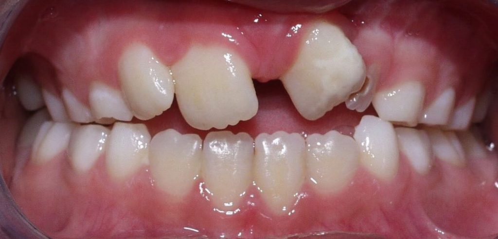
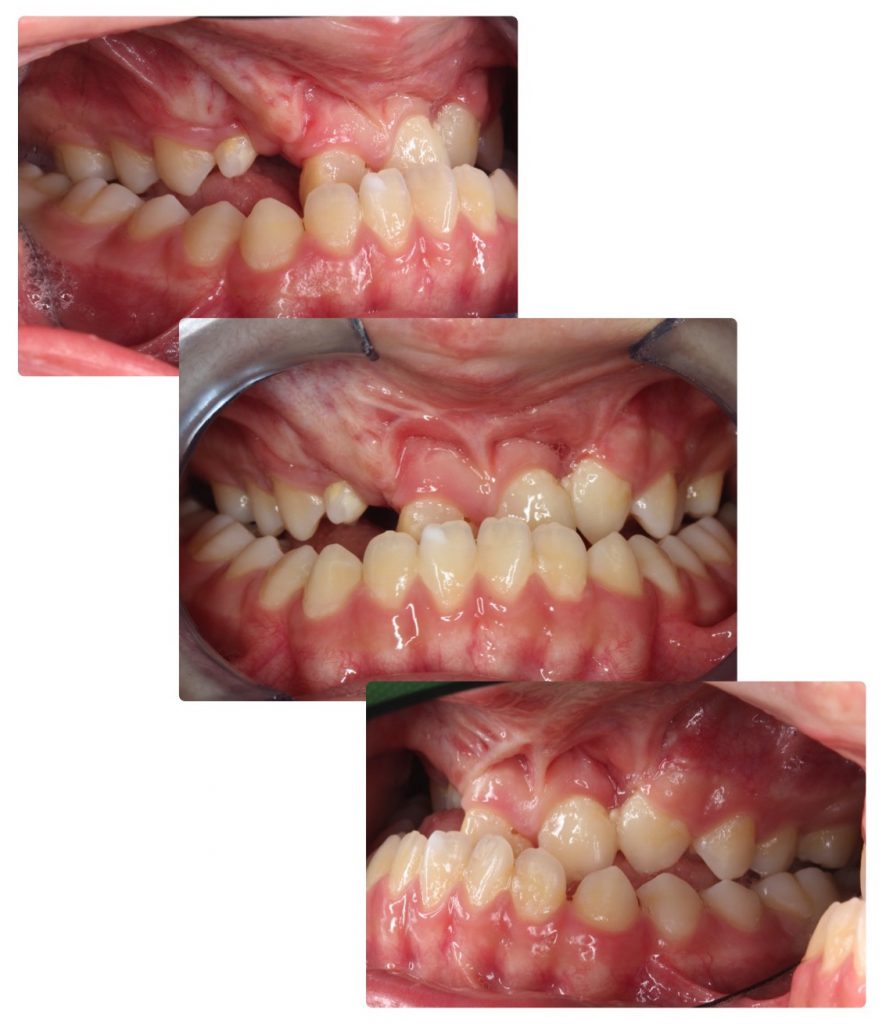
A cleft of the lip, of the alveolus (maxillary) and / or of the palate can affect the teeth in various ways. The deciduous teeth and permanent teeth can be altered in their shape, size, number and position. Most often, the teeth located in the immediate vicinity of the cleft are affected: the incisors. The alveolar cleft is located at the site of the future canine and lateral incisor.
The dental sequelae of an CLP affect the aesthetics and masticatory function of the patient. The treatment of these sequelae therefore constitutes for the patient and his/her parents an absolute priority, that the orthodontist and the maxillofacial surgeon of the team will have to actively address.
The efficacy of their treatment depends on perfect coordination and planning, taking into account the subjective needs of the patient, but also factors related to his/her growth and craniofacial development. In other words, any intervention must be proposed in view of its utility and the long-term benefits it can bring to the patient. The orthodontic treatment, which can be long, should not be started too early, so as not to tire the patient, whose cooperation will be essential during the final stages of treatment, that is towards the end of his/her growth .
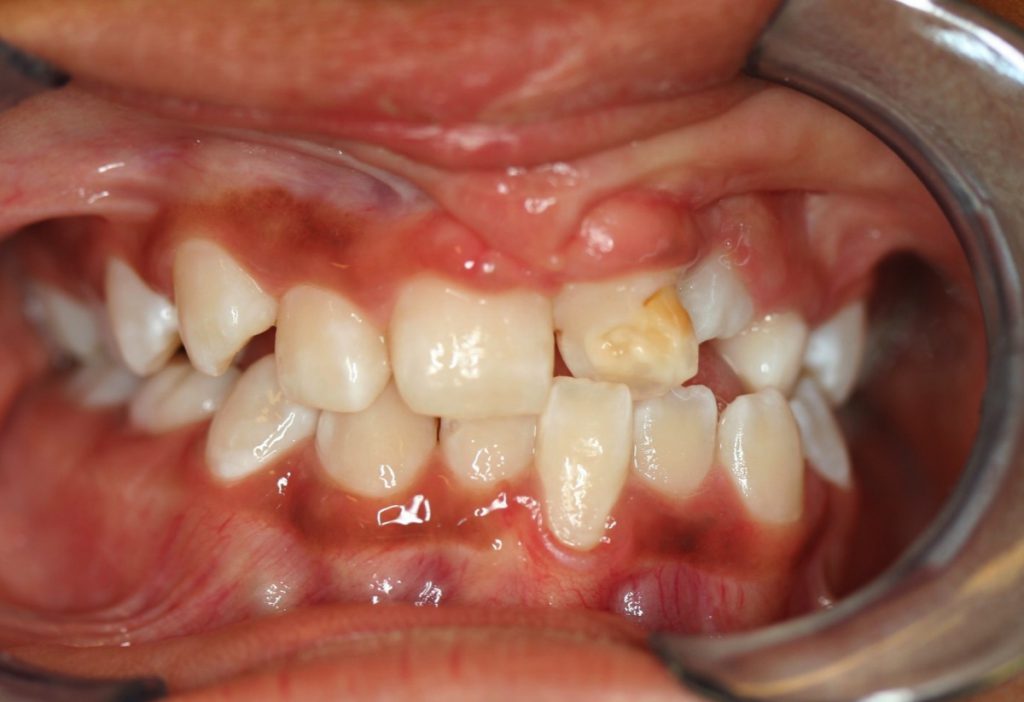
The lateral incisor may be absent (we speak of agenesis). On the contrary, it can split and there is a supernumerary tooth. Some teeth in the region of the cleft show some malformation of the crown, with for example anomalies of the mineralization or the calcification of the enamel (superficial layer of the tooth), giving an irregular aspect of the surface or white or brown spots. We speak of hypoplasia of the enamel (Fig.1).


The presence of a facial cleft disrupts the harmony of the dental arch by its adverse influence on the formation of teeth, their eruption and their subsequent position. The scars that result, inevitably, from the surgical closure of the palate, alveolus and lip, negatively influence the future growth of the upper jaw, the dental arch and, finally, the position of the teeth. .
CRANIO-FACIAL DEVELOPMENT
The malformation of the maxillary complex present at birth and the scars resulting from the first surgical operations can cause an inhibition of the growth of the upper jaw, in the three dimensions of space:
- in width (arcade too narrow); the patient will present an crossbite (upper dental arch too narrow)
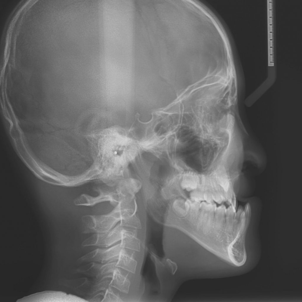
- from back to front (insufficient projection of the middle part of the face or, in other words, upper jaw remaining a little too “retruded”). It is referred to as maxillary hypoplasia or retrognathia (Fig.5 & 6): the upper jaw (whose growth is inhibited) will have insufficient projection compared to the lower jaw, whose growth is normal.
- in the vertical direction. In this case, there will be an openbite (no contact between the teeth).


Modern surgical techniques and the experience of our surgical team, however, have brought, in recent years, significant improvements in the prognosis of growth of the upper jaw in these children, essentially thanks to a significant reduction of scar tissue resulting from less traumatic surgical techniques.
Fewer scars mean better jaw growth and greater harmony of the dental arch. But the future need for orthodontic correction is still very great.
In fact, virtually all children or adolescents who have had a facial cleft will need, at one time or another, an orthodontic correction.
Georges Herzog, orthodontist consultant of the multi-disciplinary FLMP team at CHUV (Lausanne) and HUG (Geneva) contact: info@cleft-palate.com